The HHG radiation extends from 100 to 600 eV, covering the entire "water window", i.e., the spectral region below the oxygen K-shell absorption edge at about 540 eV (2.3 nm), with exceptionally high photon flux and stability in a user-friendly environment.
Exploiting these unique capabilities in a combined reflectometry and spectroscopy setup developed at MBI, a team of researchers from Thuringia, Saxony and Berlin has taken an important step toward non-destructive nanoscale imaging with soft X-rays. In the work published in Light: Science & Applications, the researchers show for the first time that high harmonics can be used for imaging in the so-called water window (approx. 2.3–4.4 nm wavelength)—a spectral range that is particularly attractive because it enables high spatial resolution and comparatively high penetration depth in conjunction with strong elemental contrast.
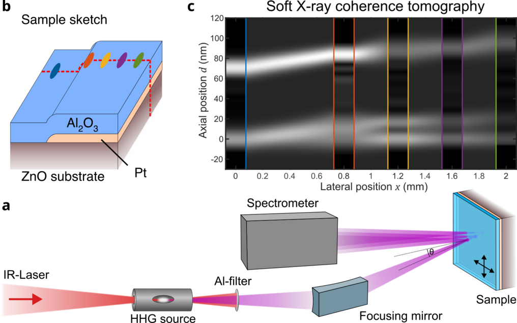
At the core of the work is a broadband method called Soft X-ray Coherence Tomography (SXCT), derived from optical coherence tomography (OCT), a standard imaging method used in medicine to obtain depth profiles, e.g., of the retina in the human eye. Using this novel technique with soft X-rays, the team explored how axial depth profiles can be obtained in reflection from solid state heterostructures and buried interfaces, structures which are of critical importance in materials science and microelectronic devices.
Until now, HHG-driven imaging of nanometer scale structures in the water window was considered hardly practical, primarily due to the limited photon yield at higher energies. The proof-of-principle-study on a test structure consisting of aluminum oxide and platinum layers now shows that the combination of a powerful, broadband HHG source (exceeding a photon flux of 10⁶ photons/eV/s on the sample) and bandwidth-efficient coherence tomography can overcome this barrier. In the SXCT approach, depth resolution in axial imaging is achieved by recording the interference signal of the soft X-ray pulses reflected from the different interfaces within a sample. By exploiting the full spectral bandwidth of the HHG source to observe the interference in the wavelength domain, the team reconstructed depth profiles with only 12 nm axial resolution in a non-destructive imaging modality. It was demonstrated that this was possible even for very weakly reflecting samples with a rough surface, thanks to the Fourier-based, lock-in-like noise suppression of the SXCT approach.
The results are obtained in a collaboration between several research organizations with contributions ranging from laser and radiation source development to tomography methodology, sample preparation and validation by electron microscopy. Partners include, among others, Friedrich Schiller University Jena and the Helmholtz Institute Jena, the Max Born Institute and TU Berlin, the Laser Institute at the University of Applied Sciences Mittweida, as well as the Leibniz Institute of Photonic Technology (IPHT) Jena.
