X-ray microscopes provide the ability to penetrate through matter and to resolve tiny features that remain hidden with visible light. Typically, X-ray images are recorded in a monochrome fashion – that is, the images appear in black and white. However, the spectroscopic X-ray response of an object – which is the direct analog of the object’s color when using visible light – contains essential information such as its elemental composition as well as the material’s chemical, electronic or magnetic state. In a study recently published in the journal Science Advances, a team of researchers from ICFO Barcelona, Max Born Institute, Technical University Berlin, Vanderbilt University and Aarhus University reported a breakthrough in high-resolution X-ray imaging with multiple colors using a lensless setup. In their experiment, the researchers investigated vanadium dioxide – a famous correlated quantum material – and clearly identified the spectroscopic fingerprints of its metallic and insulating phases with nanometer-scale spatial resolution.
Quantum materials exhibit spectacular properties like superconductivity, but are in many cases still poorly understood. In particular, spatial inhomogeneity of the materials on the nanometer-scale seems to largely influence their properties and function. In vanadium dioxide, for example, a metallic state (phase) and an insulating state can exist at the same time but at different, closely adjacent locations in the material. Key to understanding these materials is being able to observe and interpret the coexisting phases in these materials with high spatial resolution.
The imaging method used by the scientists is based on a holographic imaging approach for X-rays which was combined with sophisticated numerical post-processing of the data. Instead of imaging at one color in the X-ray spectrum, they collected a whole stack of images in a range of different X-ray wavelengths, which coincided with characteristic absorption features of the material. From the data, they then built up a hyperspectral image – that is, they were able to see the sample in full “technicolor light” rather than in black and white. In contrast to conventional X-ray microscopy methods, the approach of combining holographic imaging with numerical methods dispenses from high-resolution focusing elements for image formation. As a result, the achievable spatial resolution of this lensless approach is fundamentally only limited by the wavelength rather than by the properties of the X-ray optics. Possibly even as important in the study of functional quantum materials, the lensless setup makes room for a wide range of sample environments (for example to cool to very low temperatures or apply strong magnetic fields) and is fully compatible with time-resolved measurements, for instance, based on laser excitation of the sample.
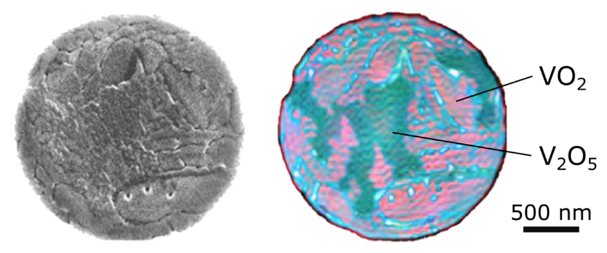
The team of scientists applied this hyperspectral x-ray imaging technique to investigate the insulator-to-metal phase transition in vanadium dioxide (VO2) upon heating the sample. While this material has been studied frequently in the past and is considered prototypical for this transition, the researchers made surprising observations. Contrary to their expectations, it became clear due to the chemical sensitivity enabled by the hyperspectral imaging that what macroscopically seemed to be pure film of VO2 turned out to have considerable amounts of V2O5 in it, distributed in small chunks of sub-micrometer size. This patchwork structure cannot be discerned in standard scanning electron microscopy images of the film and may have been overlooked in various studies of this material in the past. As vanadium oxide V2O5 lacks the characteristic phase transition, its sizeable content significantly influences the overall properties of the sample and makes the knowledge of this nanometer-scale patchwork structure crucial for the interpretation of many experiments. Regarding the insulator-to-metal phase transition, the researchers saw that the VO2 patches switch directly from the insulating to the metallic state. Previous work on this material had suggested the presence of additional intermediate phases appearing locally.
What is particularly noteworthy about the newly established hyperspectral approach to imaging with X-rays is that it is readily compatible with time-resolved experiments, hence allowing to combine spectral, temporal and spatial information. Combined with ultrashort X-ray and laser pulses, the technique promises to enable multidimensional time-resolved imaging under extreme conditions, where existing methods cannot operate. This capability is important to discover new phases in quantum materials which are elusive because they appear only for a short time or in tiny parts of a material.
